Anatomy of the Foot
Understanding the anatomy of the foot is useful when going barefoot. Knowing where all the parts are, what they look like, how they sit in relation to one another, gives a better understanding of the foot as a whole, and enables better communication between client and trimmer.
I have put together some simple diagrams to illustrate the parts of the foot and their relationships. I haven’t included ligaments, nerves or blood supply.
I have also written simple descriptions outlining each part; I have kept the descriptions short as this is intended only as a guide and I will go into more detail about function elsewhere, in the relevant section on the web site.
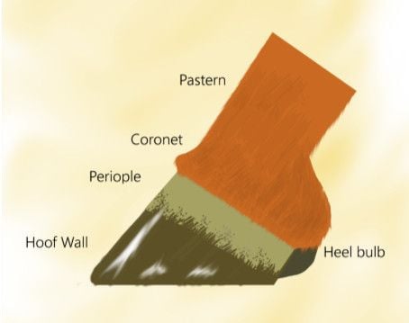
Healthy foot diagram side view
Coronet
The coronary band is one of the main sensitive tissues of the foot and is where the hoof wall forms. It is also called the coronet, this is a reference to its shape, encircling the upper part of the foot like a coronet. Its role is to supply blood to the structures of the foot.
Periople
The Perioplic Corium is responsible for producing the outer shiny layer of hoof wall. This outer layer prevents moisture loss from the inner layer of hoof wall. It also stops moisture ingress into the inner layer. The periople itself can be seen as the thin membrane that grows from the outer edge of the coronary band and down the hoof wall.
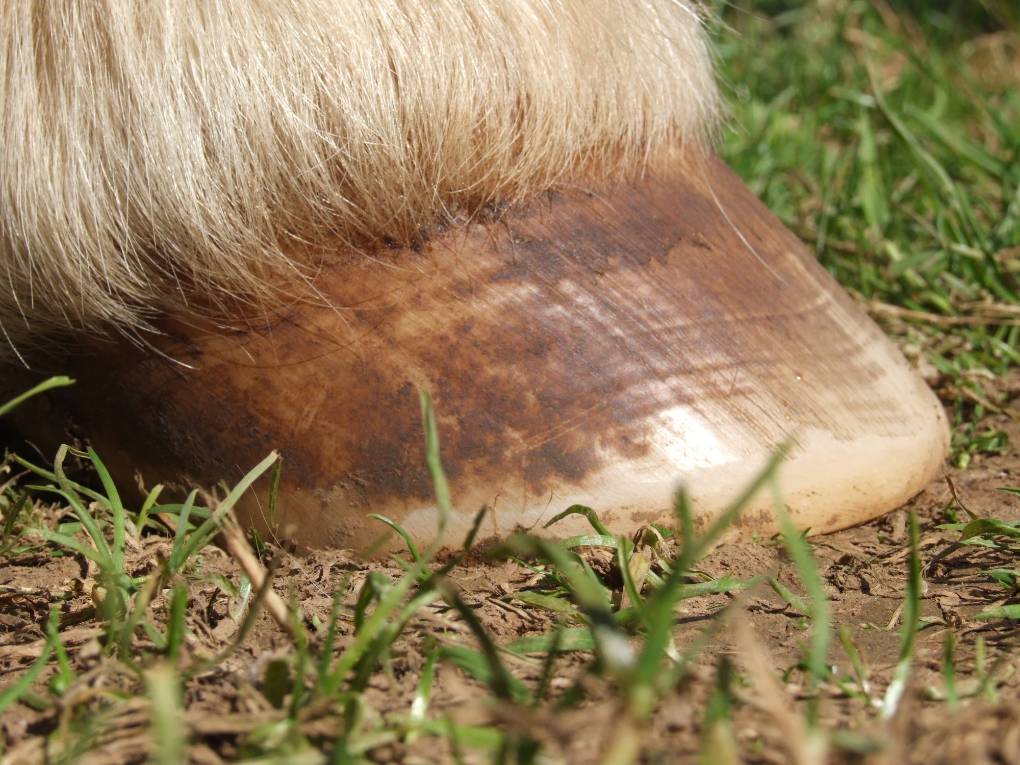
In most healthy feet the periople can be seen growing as much as a third of the way down the hoof wall.
Hoof wall
The hoof wall is initially produced at the coronet and then added to by the laminae as the wall grows, moves, down the foot. You could think of the hoof wall in three layers. The outer layer (stratum externa) the inner or middle layer, and the layer that is integrated into the epidermal lamina. However this is really like viewing the foot as separate from the horse, all these layers combine as one and for the most part have no really defining boundaries.
The hoof wall really is quite dynamic and fluid in nature.
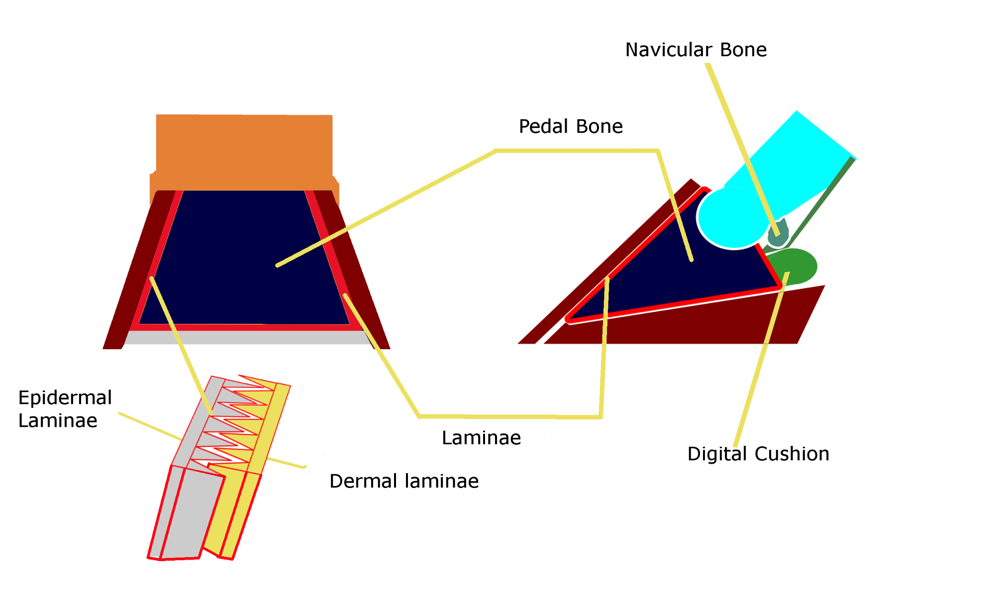
The left hand diagram shows the foot with the front part of the hoof wall removed . In the right hand diagram part of the pedal bone has been removed to show Digital Cushion and Navicular bone clearly
Laminae of the hoof wall.
Epidermal and dermal laminae attach the hoof wall to the pedal bone. The laminae are truly incredible. They provide a strong attachment of the hoof wall to the pedal bone, whilst allowing the hoof wall to grow and move down, over the pedal bone. This is no mean feat when you think about the incredible forces placed on the hoof wall when a horse moves at speed, or jumps.
The epidermal and dermal laminae are like the interlocking pages of two books. (see diagram….) This gives a large area of attachment; around one metre square of surface area in an average horse. There is a physical bond between the epidermal and dermal layers that provides extra strength. It is the breaking and remaking of these physical bonds that allows the epidermal laminae to slide over the dermal laminae as the hoof wall grows. I will write more about the laminae later as they are quite complex and many things can affect the way they function.
Pedal bone
The pedal bone or third phalanx is the last bone in the leg. It is also sometimes called the coffin bone. Both the hoof wall and sole attach to this structure. In this diagram (ref diagram) I have used a wedge shape to represent the pedal bone; I feel that this is truer to the bones actual shape. In many diagrams it is represented in cross section to allow illustration of the attachment of the deep digital flexor tendon. There is a lot to say about the pedal bone and its orientation within the hoof capsule and is something that I will expand on in other sections, most notably when discussing Navicular Syndrome.
The Navicular Bone
The distal sesamoid or navicular bone is the small bone that the deep digital flexor tendon runs over before attaching to the Pedal Bone
Digital Cushion
The digital cushion and its surrounding fibrocartilage is one of the most important structures of the foot but the least talked about. The primary function of the digital cushion is to absorb shock and reduce concussion to the limb. It also plays a major role in circulating blood through the foot.
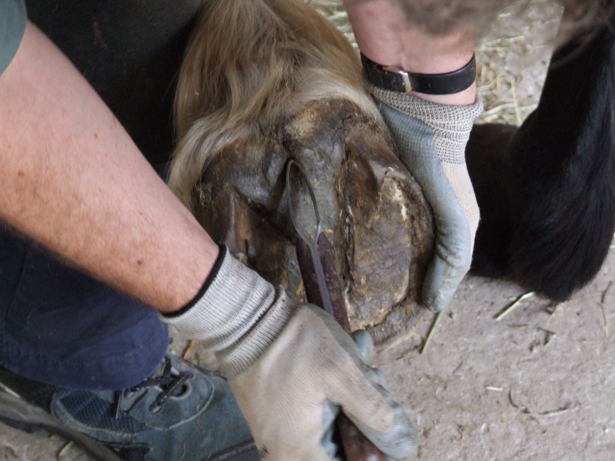
Heel Bars
The heel bars are part of the hoof wall that wraps around the back of the foot and form the inner part of the heel. They also form part of the collateral groove that runs around the frog. This groove should be deeper at the heel and become shallower at the apex of the frog
The frog
The frog is a mass of soft keratinised tissue that should be in contact with the ground and is meant to be weight bearing.
The central sulcus should be a shallow bowl like scoop within the surface of the frog, not a split that divides the heel bulbs. The importance of a healthy frog is widely underestimated.
Sole and Sole Corium
Sole corium is a vascular structure, that manufactures sole. The sole is attached to the bottom of the foot in very much the same way as the hoof wall. The keratin produced by the sole corium is similar to the hoof wall but acts differently. The keratin that makes up the sole requires load to pack it down and become hard and wear resistant. If sole is produced in areas of little or no loading it is quite soft and flaky.
Ungular Cartilage
Also known as the collateral cartilage, supports the coffin joint. It also plays a role in the dissipation of energy when blood is drawn through it.
The cartilage itself can ossify and become less effective.This is often referred to as sidebone.